
Collagen Imaging Using Fluorescent Peptides
Article Sidebar
Main Article Content
Abstract
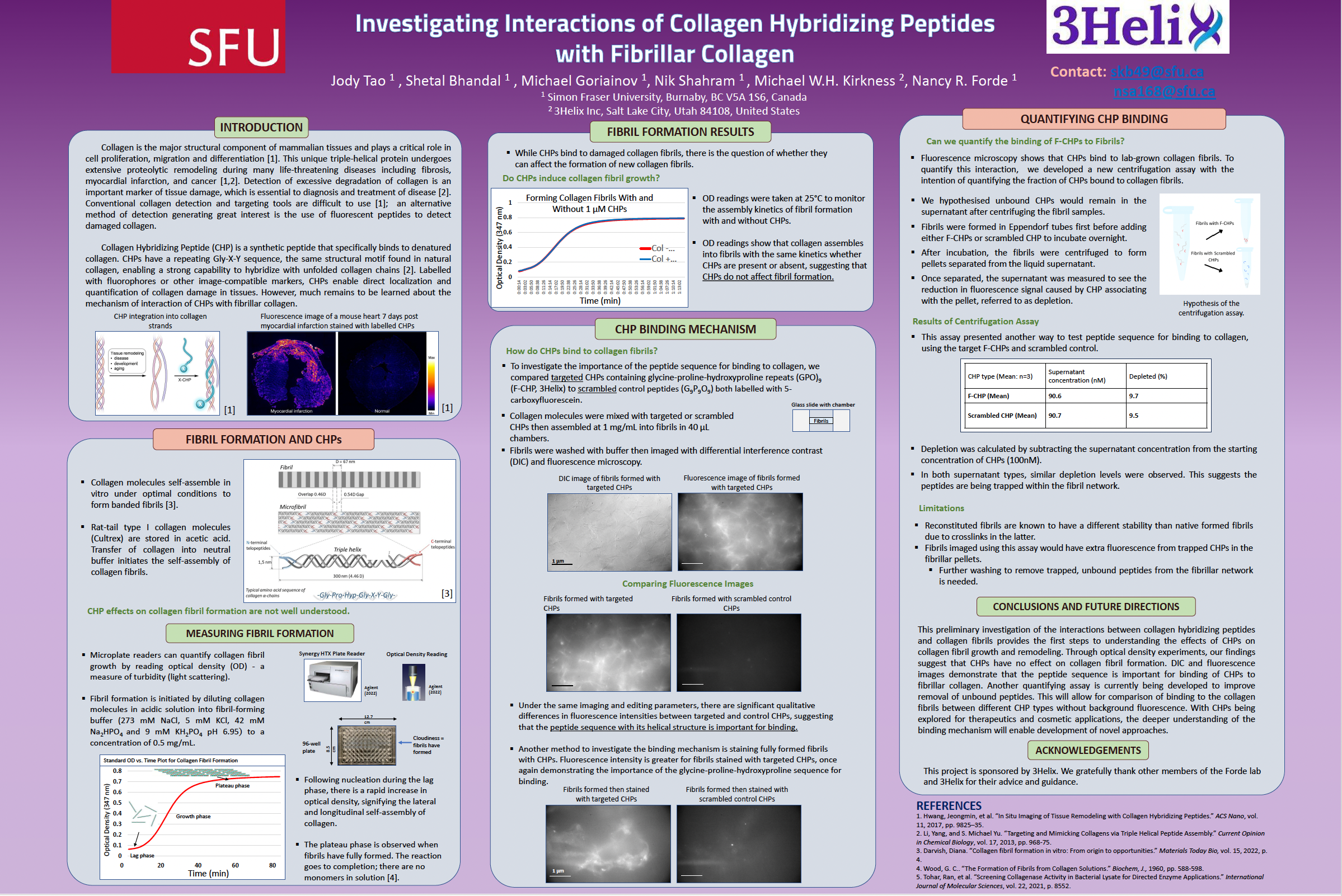
Collagen is the major structural component of mammalian tissues and plays a critical role in cell proliferation, migration and differentiation. This unique triple-helical protein undergoes extensive remodeling during many life-threatening diseases including fibrosis, myocardial infarction, and cancer. Detection of excessive degradation of collagen is an important marker of tissue damage, which is essential to the diagnosis and treatment of disease. Conventional collagen detection and targeting tools are difficult to use; an alternative method of detection generating great interest is the use of Collagen Hybridizing Peptides (CHPs) which are fluorescently labelled peptides that detect damaged collagen. CHPs have a repeating Gly-X-Y sequence, the same motif found in natural collagen, which provides a strong affinity to hybridize with unfolded collagen chains. CHPs enable direct localization of collagen damage in tissues. However, much remains to be learned about the mechanisms by which these peptides interact with fibrillar collagen, as well as with gelatin networks. Here, our experiments with fluorescence microscopy demonstrate that CHPs have some affinity for collagen fibrils. They show the importance of the repeating Gly-X-Y sequence, through comparison with experiments using peptides with this sequence order scrambled. We are extending this work to quantify the binding of CHPs to gelatin networks, which contain much higher amounts of damaged collagen. These assays lay the groundwork for future experiments that provide additional insight into the binding mechanism of CHPs.
Article Details

This work is licensed under a Creative Commons Attribution-NonCommercial-NoDerivatives 4.0 International License.